
“This could be the beginning of the end for painful biopsies,” the patch with millions of nanoneedles

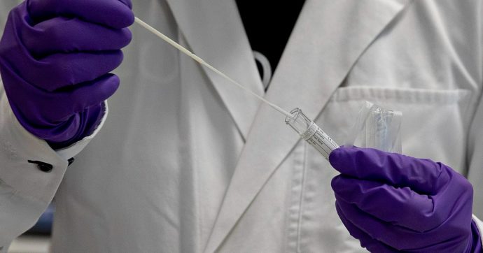
A patch containing tens of millions of microscopic nanoneedles could soon replace traditional biopsies . Developed by researchers at King's College London, the innovative patch offers a painless, less invasive alternative for millions of patients worldwide who undergo biopsies each year to detect and monitor diseases such as cancer and Alzheimer's. The findings were published in the journal Nature Nanotechnology .
Technology – Biopsies are among the most common diagnostic procedures in the world, performed millions of times each year to detect disease. However, they are invasive, can cause pain and complications, and can discourage patients from seeking early diagnosis or follow-up tests. Traditional biopsies also involve removing small pieces of tissue, limiting the frequency and accuracy with which doctors can examine diseased organs such as the brain.
Now, scientists at King’s College London have developed a nanoneedle patch that painlessly collects molecular information from tissue, without removing or damaging it. This could allow healthcare professionals to monitor disease in real time and perform multiple repeatable tests on the same area, which is not possible with standard biopsies. Because the nanoneedles are 1,000 times thinner than a human hair and do not remove tissue, they do not cause pain or damage, making the procedure less painful for patients than standard biopsies.
The utility – This could mean earlier diagnosis and more regular monitoring, transforming the way diseases are monitored and treated. “We’ve been working on nanoneedles for twelve years, but this is our most exciting development yet,” says Ciro Chiappini , who heads the research. “It opens up a world of possibilities for people with brain tumors , Alzheimer’s , and for the advancement of personalized medicine. It will allow scientists – and eventually doctors – to study diseases in real time like never before,” he adds.
The patch is covered with tens of millions of nanoneedles. In preclinical studies, the research team applied the patch to brain tumor tissue taken from human biopsies and mouse models. The nanoneedles extracted molecular “fingerprints” — including lipids, proteins, and mRNA — from the cells, without removing or damaging the tissue. The tissue fingerprint is then analyzed using mass spectrometry and artificial intelligence, providing healthcare teams with detailed information about the presence of a tumor, its response to treatment, and the progression of the disease at the cellular level. “This approach provides multidimensional molecular information from different cell types within the same tissue,” Chiappini explains.
The breakthrough – “Traditional biopsies simply can’t do this. And because the process doesn’t destroy tissue, we can sample the same tissue multiple times, which was previously impossible,” he adds. The technology could be used in neurosurgery to help surgeons make faster, more accurate decisions. For example, by applying the patch to a suspicious area, results could be obtained within 20 minutes and could guide real-time decisions about removing cancerous tissue. Made using the same manufacturing techniques as computer chips, the nanoneedles could be integrated into common medical devices such as bandages, endoscopes and contact lenses . “This could be the beginning of the end of painful biopsies,” Chiappini says. “Our technology opens up new ways to diagnose and monitor disease safely and painlessly, helping doctors and patients make better, faster decisions,” he adds. This breakthrough was made possible by the close collaboration between nanoengineering, clinical oncology, cell biology, and artificial intelligence: each field contributed essential tools and perspectives that, together, unlocked a new approach to noninvasive diagnostics.
Valentina Arcovio
Il Fatto Quotidiano


